When a person is diagnosed with cancer, one of the first and most critical questions is: Where is the cancer, and how far has it spread? This isn’t just about finding a tumor. It’s about understanding whether it’s still localized, if it’s reached lymph nodes, or if it’s jumped to distant organs like the liver, lungs, or bones. The answer determines everything - whether someone gets surgery, chemotherapy, radiation, or a combination. And that answer comes from imaging. Not just any imaging, but the right kind at the right time.
Three technologies dominate modern cancer staging: PET-CT, MRI, and the newer hybrid PET-MRI. Each has strengths, weaknesses, and situations where it shines. Choosing one over the other isn’t about which is "better." It’s about which one gives the clearest picture for your cancer, at this stage, with your body.
PET-CT: The Workhorse of Cancer Staging
PET-CT became the standard in the early 2000s, and for good reason. It combines two scans into one. The PET part uses a radioactive sugar tracer - usually 18F-FDG - that cancer cells gobble up because they’re hungry for energy. The CT part gives a detailed anatomical map. Together, they show not just where a tumor is, but whether it’s active.
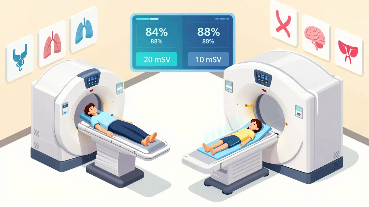
For cancers like lymphoma, lung cancer, or melanoma, PET-CT is often the first imaging test after diagnosis. It can find tiny metastases that CT or MRI alone might miss. A 2023 meta-analysis showed PET-CT correctly identified lymph node involvement in non-small cell lung cancer with 84% accuracy. It’s fast - a full-body scan takes 15 to 20 minutes. It’s widely available. Most hospitals have one.
But it has limits. Not all cancers use sugar aggressively. Some prostate cancers, low-grade tumors, or certain types of brain cancer barely light up on PET. And because it uses radiation - typically 10 to 25 millisieverts per scan - repeated scans aren’t ideal, especially for younger patients or those needing long-term monitoring.
MRI: Seeing the Invisible
MRI doesn’t use radiation. Instead, it uses powerful magnets and radio waves to create images based on how water behaves in different tissues. This gives it unmatched detail in soft tissues - the brain, spinal cord, liver, prostate, and pelvic organs.
For prostate cancer, MRI is now the go-to before biopsy. A 2022 study found it detected cancer with 75% accuracy, outperforming PSMA PET-CT in some cases. In breast cancer, especially after chemotherapy, MRI can show whether the tumor is truly shrinking or just scar tissue. And for liver tumors, MRI with contrast agents can distinguish between benign growths and metastases better than any other tool.
But MRI has downsides. It’s slow. A detailed scan can take 30 to 60 minutes. You have to lie perfectly still. People with metal implants - pacemakers, cochlear implants, some surgical clips - can’t have it. And MRI alone doesn’t tell you if a tumor is metabolically active. A dark spot could be cancer… or just inflammation.
PET-MRI: The High-Tech Hybrid
PET-MRI, introduced commercially in 2011, merges the metabolic power of PET with the soft-tissue precision of MRI - all in one machine. It’s not just a side-by-side scan. It’s simultaneous. That means the data aligns perfectly. No guesswork about whether a hot spot on PET lines up with a lesion on MRI.
This is where PET-MRI truly changes the game. In brain tumors, it can tell the difference between recurring cancer and radiation damage - something MRI alone struggles with. Studies show PET-MRI gets it right 85-90% of the time, compared to 70-80% for MRI alone. For pediatric cancers, where minimizing radiation is critical, PET-MRI cuts exposure by nearly half compared to PET-CT. In pelvic cancers - cervical, rectal, or ovarian - it gives surgeons a 3D map of tumor spread that changes treatment plans in nearly half of cases.
But it’s not perfect. The scan takes longer - 45 to 60 minutes. Motion from breathing or bowel movement can blur the images. It’s expensive. A single PET-MRI scan costs $2,500 to $3,500 in the U.S., nearly double the cost of PET-CT. And not every hospital has one. Only 78% of PET-MRI installations are in academic centers with research programs. Many community hospitals still rely on PET-CT because it’s faster, cheaper, and familiar.
Which One Is Right for You?
There’s no universal answer. The best imaging choice depends on your cancer type, location, and treatment goals.
- For lung, lymphoma, or melanoma: PET-CT remains the standard. It’s fast, proven, and shows spread across the whole body.
- For prostate, liver, or pelvic cancers: MRI or PET-MRI is often superior. They show local invasion and subtle changes that PET-CT misses.
- For brain tumors: PET-MRI is the gold standard. It’s the only way to reliably tell if a growing area is tumor recurrence or treatment-related scarring.
- For children or young adults: PET-MRI is preferred when available. Reducing radiation over a lifetime matters.
- For monitoring treatment response: PET-CT is still used for quick checks. But if a scan shows unclear results, switching to MRI or PET-MRI can clarify.
Even experts don’t agree on a one-size-fits-all approach. Dr. Richard L. Wahl from Johns Hopkins says PET-MRI is "particularly valuable for pelvic malignancies and CNS tumors," but adds that PET-CT remains the "workhorse." Dr. Hedvig Hricak from Memorial Sloan Kettering puts it plainly: "The choice must be personalized."

Real-World Challenges
It’s not just about what’s on the screen. Practical issues shape what you get.
One major hurdle is time. A PET-MRI scan can take over an hour. For someone in pain, with difficulty lying still, or with anxiety, that’s a challenge. Technologists report motion artifacts - blurry images from movement - are common in abdominal PET-MRI scans. Some centers have started using sedation for these cases.
Cost and access are bigger barriers. A PET-CT machine costs $1.8-2.5 million. A PET-MRI? $3-4.2 million. That’s not just the machine. It’s a shielded room, specialized staff, and ongoing maintenance. Only 45% of U.S. cancer centers have PET-MRI. Reimbursement is inconsistent. Some insurance plans still don’t cover it unless PET-CT was inconclusive.
Training matters too. Interpreting PET-MRI requires understanding both nuclear medicine and advanced MRI sequences. Radiologists need an extra 3-6 months of training beyond what’s needed for PET-CT. A 2022 survey found that 63% of sites reported difficulties with attenuation correction - a technical glitch that can distort PET images if not handled properly.
The Future: AI, New Tracers, and Personalized Imaging
The next wave is already here. In January 2024, Siemens Healthineers got FDA clearance for its BioMatrix 600 PET-MRI system - a machine that can scan the whole body in just six minutes. That cuts scan time by 50%, making it more practical for busy clinics.
New tracers are changing the game too. PSMA PET-CT, approved in 2020, is now standard for prostate cancer. It’s far more sensitive than older tracers. In 2024, ASCO guidelines began recommending imaging choices based on tumor biology - not just location. A HER2-positive breast cancer may respond differently to PET-CT than a triple-negative one. This is the future: imaging tailored to your cancer’s genetic fingerprint.
Artificial intelligence is starting to analyze patterns in these scans that humans miss. Radiomics - extracting hundreds of data points from an image - is being tested in clinical trials to predict whether a tumor will respond to therapy before treatment even begins. The NCI’s PREDICT trial is tracking over 1,200 patients using AI-assisted PET-MRI to guide personalized treatment decisions.
By 2035, experts predict PET-MRI will make up 25-30% of oncologic imaging in academic centers. But PET-CT won’t disappear. It’s too practical, too reliable, too widespread. The future isn’t one technology replacing another. It’s using the right tool for the right job - and knowing when to switch.
Is PET-CT or MRI better for cancer staging?
There’s no single answer. PET-CT is best for whole-body surveys - finding spread in lung, lymphoma, or melanoma. MRI excels at detailed views of soft tissues like the brain, prostate, liver, or pelvic organs. PET-MRI combines both and is superior for complex cases, like brain tumors or pediatric cancers. The choice depends on cancer type, location, and treatment goals.
Does MRI detect cancer better than PET-CT?
It depends. MRI gives better anatomical detail in soft tissues, so it’s better for spotting small tumors in the prostate, liver, or brain. PET-CT shows metabolic activity, so it’s better for finding hidden spread - like a tiny lymph node tumor that looks normal on MRI. In breast cancer, PET-CT has higher specificity for early treatment response. In prostate cancer, MRI is often more accurate for initial detection. They’re complementary, not competitive.
Why is PET-MRI so expensive?
PET-MRI machines cost $3-4.2 million - nearly double the price of PET-CT. They require a magnetically shielded room ($150,000-$250,000), specialized physics support, and highly trained staff. The scan itself takes longer, reducing patient throughput. Insurance reimbursement is inconsistent, and many centers can’t justify the cost unless they’re doing high-volume research or treating complex cases like pediatric cancer or recurrent brain tumors.
Can PET-MRI replace PET-CT entirely?
No. PET-CT remains the workhorse for most cancers because it’s faster, cheaper, widely available, and highly effective for whole-body staging. PET-MRI is a specialized tool that outperforms PET-CT in specific situations - like brain tumors, pelvic cancers, or when radiation reduction is critical. The trend is toward using each modality where it’s strongest, not replacing one with the other.
Is radiation from PET-CT dangerous?
A single PET-CT scan delivers 10-25 millisieverts of radiation - roughly 3-8 times a standard chest X-ray. While the risk from one scan is low, repeated scans over time increase cumulative exposure. For younger patients or those needing long-term monitoring, PET-MRI is preferred because it cuts radiation by about 50%. For most adults with advanced cancer, the benefit of accurate staging outweighs the small long-term risk.
rakesh sabharwal
March 14, 2026 AT 12:24And let's not pretend reimbursement policies are about patient care - they're about billing codes and institutional inertia. The entire system is a Rube Goldberg machine built on ego, not evidence.
Aaron Leib
March 15, 2026 AT 09:44What matters most is matching the tool to the clinical question. PET-CT for whole-body spread. MRI for local detail. PET-MRI when precision changes management. Simple as that.
Dylan Patrick
March 16, 2026 AT 08:41That’s the reality for so many patients. No one talks about the human cost of these "gold standard" scans. We fixate on tech specs while people suffer through them. Just sayin’.
Kathy Leslie
March 17, 2026 AT 06:10But we still save lives. Every day. With what we’ve got. The real hero isn’t the machine - it’s the radiologist who knows when to trust the image and when to trust the patient.
Amisha Patel
March 17, 2026 AT 22:16Elsa Rodriguez
March 19, 2026 AT 19:01Serena Petrie
March 20, 2026 AT 05:16Buddy Nataatmadja
March 21, 2026 AT 02:51Technology doesn’t heal. People do.
mir yasir
March 23, 2026 AT 00:27The reliance on multimodal imaging is not an advancement - it is an epistemic indulgence.
Stephanie Paluch
March 24, 2026 AT 00:40They did MRI after chemo and I thought it was scar tissue… turns out it was still cancer. I didn’t know MRI could see that. Thank you for explaining this so clearly. I feel less alone now 💕
tynece roberts
March 24, 2026 AT 10:02so i read this whole thing and now i get it kinda??
but like… why is it so expensive?? like i get the machine costs a lot but like… can’t we just… make cheaper ones??
also i think the part about kids and radiation is super important. my cousin had 3 pet-cts by age 12. that’s wild. i didn’t even know that was a thing.